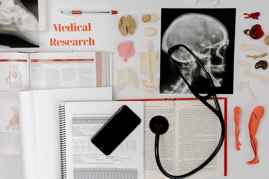
This manual, Pearson’s new 10th edition (ISBN: 1292026375, 9781292026374), provides a comprehensive, hands-on exploration of human body structures and their functions․
It emphasizes the crucial link between anatomical form and physiological processes, utilizing extensive instructor support for effective lab quizzing․
Purpose of the Lab Manual
The primary goal of this Human Anatomy & Physiology Laboratory Manual, specifically the main version, is to supplement and enhance the learning experience from the corresponding lecture course․
It aims to provide students with practical, hands-on experience in identifying and understanding the structures of the human body․ This is achieved through detailed exercises and activities, fostering a deeper comprehension of anatomical relationships and physiological mechanisms․
Furthermore, the manual supports the development of essential laboratory skills, including microscopy and dissection, while reinforcing the vital connection between structure and function․
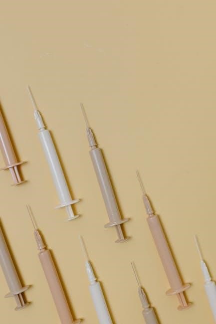
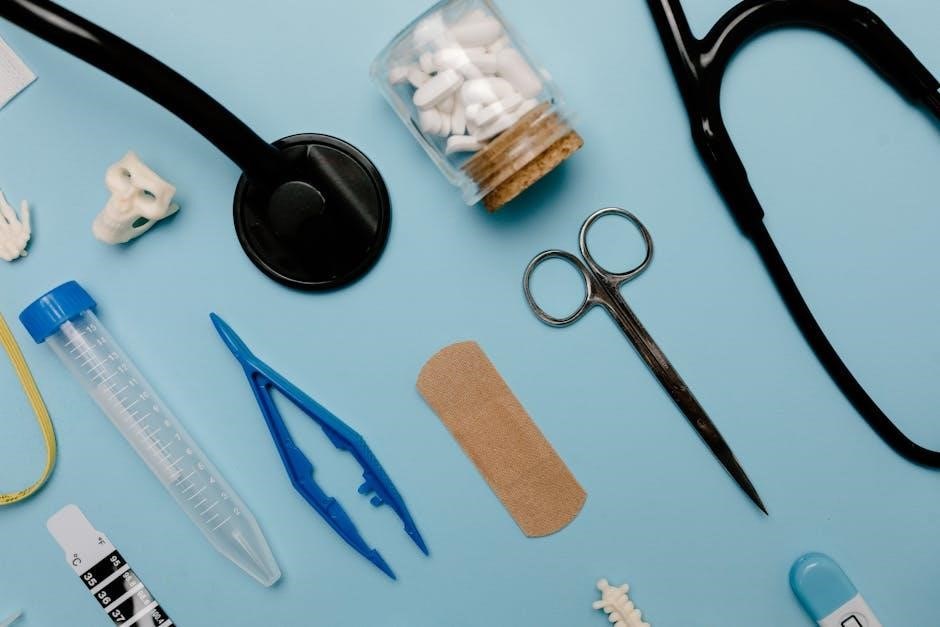
Safety Procedures in the Lab
Prioritizing safety is paramount within the anatomy and physiology laboratory environment․ Students must adhere to all instructor guidelines and established protocols at all times․
Proper handling and disposal of specimens, chemicals, and sharp instruments are crucial to prevent injury and contamination․ Always wear appropriate personal protective equipment (PPE), including gloves and eye protection․
Maintain a clean and organized workspace, and immediately report any accidents or spills to the instructor․ Familiarize yourself with emergency procedures and the location of safety equipment․ Respectful conduct and responsible behavior are essential for a safe learning environment․
Microscopy and Histology
This section details microscope usage (Chapter 2) and slide preparation, focusing on identifying epithelial and connective tissues – key components of histological study․
Using the Microscope
Mastering microscopy is fundamental to understanding anatomy and physiology․ This lab manual section (Chapter 2) provides detailed instructions on proper microscope operation, including component identification and correct usage․
Students will learn techniques for achieving optimal image clarity through focusing adjustments and magnification control․ Essential skills covered include preparing wet mounts and dry mounts for observation․
Furthermore, the manual emphasizes the importance of understanding magnification power and resolution, enabling accurate visualization of cellular structures and tissue types․ Proper handling and maintenance of the microscope are also stressed․
Preparing Microscope Slides
Effective slide preparation is crucial for clear microscopic observation․ This section details techniques for creating both wet mount and dry mount slides, essential for visualizing various specimens․
Students will learn proper methods for specimen collection, mounting, and staining, ensuring optimal contrast and detail․ Emphasis is placed on minimizing artifacts and preventing sample damage during preparation․
The manual guides users through selecting appropriate stains to highlight specific cellular structures, enhancing their visibility under the microscope․ Proper labeling and slide handling procedures are also thoroughly explained․
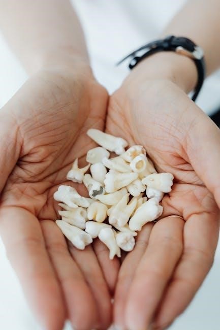
Epithelial Tissue Identification
This section focuses on mastering the identification of diverse epithelial tissue types, a fundamental skill in histology․ Students will learn to differentiate tissues based on cell shape – squamous, cuboidal, and columnar – and layering – simple, stratified, and pseudostratified․
Key characteristics, such as the presence of cilia, microvilli, and specialized junctions, will be emphasized․ The manual provides detailed microscopic images and descriptive text to aid in accurate identification․
Practical exercises will challenge students to distinguish between various epithelial tissues found in different organs, linking structure to specific functions within the body․
Connective Tissue Identification
This lab segment centers on recognizing the diverse array of connective tissues, crucial for support, protection, and integration throughout the body․ Students will differentiate tissues based on their matrix composition – collagen, elastic fibers, and ground substance – and cell types․
Emphasis will be placed on identifying hyaline cartilage, elastic cartilage, bone, blood, and various types of connective tissue proper, like adipose and dense connective tissue․
Microscopic observation and comparative analysis will be key, correlating structural features with specific functions in different anatomical locations․
The Human Cell
This section investigates the fundamental unit of life, exploring cell structure and function․ Emphasis is on relating microscopic anatomy to physiological processes within the human body․
Cell Structure and Function
This lab component delves into the intricate world of cellular biology, examining the key structures that define cell life․ Students will investigate organelles – the nucleus, mitochondria, ribosomes, and more – and their specific roles in maintaining cellular homeostasis․
The focus is on understanding how these structures contribute to overall cell function, including protein synthesis, energy production, and waste removal․ Microscopic observation and detailed analysis will reinforce the relationship between structure and function, a cornerstone of anatomy and physiology․
Emphasis will be placed on how cellular dysfunction leads to disease states․
Cell Transport Mechanisms
This section of the lab explores how substances move across the cell membrane, vital for maintaining cellular equilibrium․ Students will investigate passive transport – diffusion, osmosis, and facilitated diffusion – observing these processes firsthand through experiments․
Active transport mechanisms, requiring energy expenditure, will also be examined, including endocytosis and exocytosis․ The lab emphasizes the importance of concentration gradients and membrane permeability in regulating transport․
Understanding these mechanisms is crucial for comprehending physiological processes like nutrient absorption and waste elimination․
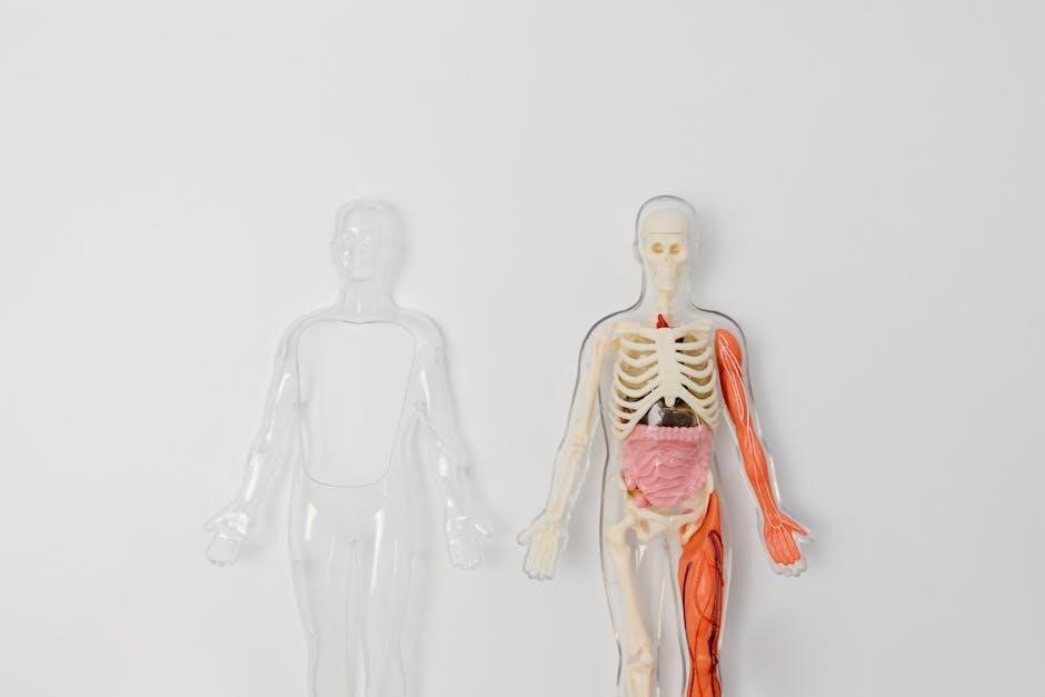
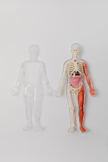
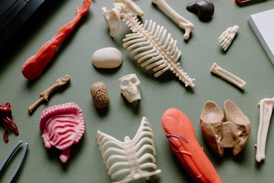
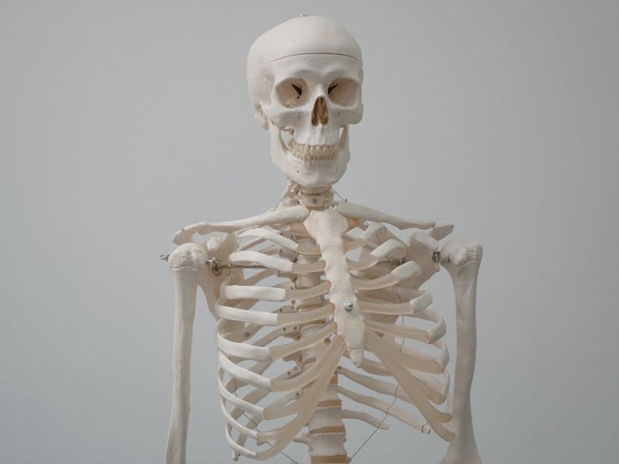
Skeletal System
This lab focuses on bone structure classification, and the axial and appendicular skeletons․ Students will identify bones and explore their relationships to movement and support․
Bone Structure and Classification
This section delves into the intricate composition of bone tissue, examining both compact and spongy bone․ Students will learn to differentiate between various bone cell types – osteoblasts, osteocytes, and osteoclasts – and understand their roles in bone remodeling․
Furthermore, the lab explores bone classification based on shape: long, short, flat, irregular, and sesamoid․ Practical exercises involve identifying these bone types and analyzing their structural features․ Understanding these classifications is crucial for comprehending bone function and biomechanics within the skeletal system․
The manual provides detailed illustrations and exercises to reinforce these concepts․
Axial Skeleton
This lab focuses on the central supporting structure of the body – the axial skeleton․ Students will identify and analyze the bones comprising the skull, vertebral column, and thoracic cage․ Detailed examination of cranial and facial bones, including sutures and foramina, will be conducted․
The vertebral column, with its regional divisions (cervical, thoracic, lumbar, sacral, coccygeal), will be studied, emphasizing vertebral structure and curvature․ Ribs and the sternum, forming the thoracic cage, will also be explored․ Practical exercises involve bone articulation and functional relationships․
The manual aids in understanding axial skeleton protection․
Appendicular Skeleton
This lab investigates the bones of the limbs and their girdles – the appendicular skeleton․ Students will identify and analyze the bones of the pectoral girdle (clavicle and scapula), upper limb (humerus, radius, ulna, carpals, metacarpals, phalanges), pelvic girdle (ilium, ischium, pubis), and lower limb (femur, tibia, fibula, tarsals, metatarsals, phalanges)․
Detailed examination of bone markings and articulation points will be performed․ Practical exercises will focus on limb movements and the skeletal support for muscle attachments․ The manual emphasizes the functional relationship between bone structure and locomotion․
Understanding the appendicular skeleton’s role in movement is key․

Muscular System
This section explores muscle tissue types, focusing on skeletal, smooth, and cardiac muscle․ Labs will examine muscle contraction mechanisms and the relationship between structure and function․
Muscle Tissue Types
This lab component delves into the three primary muscle tissue types: skeletal, smooth, and cardiac․ Skeletal muscle, responsible for voluntary movements, will be examined microscopically, noting its striated appearance and multinucleated fibers․ Smooth muscle, found in the walls of internal organs, will be contrasted, highlighting its non-striated nature and role in involuntary functions like digestion․
Cardiac muscle, unique to the heart, will be studied for its striated pattern, branching fibers, and intercalated discs – crucial for coordinated contractions․ Students will learn to differentiate these tissues based on microscopic characteristics and functional roles, understanding how each contributes to overall body physiology․
Muscle Contraction
This section explores the physiological mechanisms driving muscle contraction․ Students will investigate the sliding filament theory, examining the roles of actin, myosin, and calcium ions in the contraction process․ Lab activities may include modeling muscle fiber shortening and analyzing the effects of varying stimuli on contraction strength․
Emphasis will be placed on the neuromuscular junction and the transmission of nerve impulses to muscle fibers; Understanding the energy requirements for muscle contraction, including ATP utilization, is crucial․ The lab will reinforce the relationship between muscle physiology and overall body movement and function․

Nervous System
This section details brain anatomy, the spinal cord, and peripheral nerves․ Labs focus on identifying structures and understanding neural pathways for signal transmission and response․
Brain Anatomy
The laboratory exercises pertaining to brain anatomy provide a detailed exploration of the central nervous system’s control center․ Students will identify major brain regions – cerebrum, cerebellum, and brainstem – and their associated lobes․
Dissections, or models, will be utilized to visualize gyri, sulci, and key functional areas like the motor cortex and sensory areas․ Emphasis is placed on understanding the relationship between specific brain structures and their roles in higher-level functions, including cognition, emotion, and motor control․
Furthermore, students will examine the protective layers of the meninges and the ventricular system, crucial for cerebrospinal fluid circulation and brain protection․
Spinal Cord and Peripheral Nerves
This section of the lab manual focuses on the spinal cord’s structure and function as a vital communication pathway․ Students will identify the cord’s external features – dorsal and ventral horns, and the central canal – through models and diagrams․
The lab will explore the organization of white and gray matter, and the role of nerve tracts in transmitting sensory and motor signals․ Peripheral nerves, including cranial and spinal nerves, will be examined, emphasizing their distribution and innervation patterns․
Understanding the relationship between nerve structure and function is key, alongside exploring reflex arcs and their clinical significance․
Cardiovascular System
This lab investigates the heart’s anatomy and blood vessel structure․ Students will trace blood flow, identify chambers, valves, and major arteries/veins, enhancing understanding of circulation․
Heart Anatomy
This section focuses on detailed examination of the heart’s internal and external structures․ Students will identify the four chambers – right and left atria, and right and left ventricles – and trace the pathway of blood through them․
Key features like the atrioventricular valves (tricuspid and mitral) and semilunar valves (pulmonary and aortic) will be located and their functions explained․ The lab also covers the coronary arteries and veins, crucial for heart muscle perfusion, alongside the pericardium and its layers․
Dissection or models will aid in visualizing the myocardium, chordae tendineae, and papillary muscles, solidifying understanding of cardiac mechanics and structure-function relationships․
Blood Vessels
This lab explores the structure and function of arteries, veins, and capillaries, the vital network transporting blood throughout the body․ Students will differentiate between these vessel types based on their wall thickness, lumen size, and valve presence․
Microscopic examination of vessel cross-sections will reveal the layers – tunica intima, media, and adventitia – and their composition․ The lab also covers the systemic and pulmonary circuits, tracing blood flow and understanding blood pressure regulation․
Activities may include identifying major arteries and veins on anatomical models, and analyzing the relationship between vessel structure and its role in circulation․
Respiratory System
The lab focuses on lung anatomy and gas exchange mechanisms, examining structures like the trachea, bronchi, and alveoli for efficient oxygen and carbon dioxide transfer․
Lung Anatomy
This section of the lab manual meticulously details the anatomical structures of the respiratory system, with a primary focus on the lungs․ Students will investigate the branching pattern of the trachea into the primary bronchi, and further into the bronchioles, culminating in the alveoli – the sites of gas exchange․
Dissections and microscopic observations will highlight the pleural membranes surrounding each lung, and the role of the diaphragm in ventilation․ Emphasis is placed on understanding the structural adaptations that maximize surface area for efficient oxygen uptake and carbon dioxide removal, crucial for maintaining homeostasis․
Detailed diagrams and interactive exercises will reinforce comprehension of the lung’s complex architecture․
Gas Exchange
This lab module delves into the physiological mechanisms governing gas exchange within the lungs and at the cellular level․ Students will explore the principles of diffusion, focusing on oxygen and carbon dioxide movement across the alveolar-capillary membrane․
Experiments will demonstrate the impact of partial pressure gradients and surface area on the rate of gas exchange․ Investigations will also cover the role of hemoglobin in oxygen transport and the factors influencing its affinity for oxygen․
Understanding these processes is fundamental to comprehending respiratory function and its regulation, vital for maintaining systemic oxygen delivery․
Digestive System
This section examines digestive organs and their functions, tracing the pathway of food processing․ Labs will explore mechanical and chemical digestion, absorption, and elimination processes․
Digestive Organs and Functions
This lab meticulously investigates the anatomy and physiological roles of each digestive organ, starting with the mouth and progressing through the esophagus, stomach, small intestine, and large intestine․ Students will analyze the unique structural adaptations of each organ, correlating them with specific digestive functions like mechanical breakdown, enzymatic digestion, nutrient absorption, and waste compaction․
Emphasis is placed on understanding the accessory organs – the liver, gallbladder, and pancreas – and their contributions to digestion through bile and enzyme production․ Dissection and microscopic observation will enhance comprehension of tissue organization and functional specialization within the digestive tract․
Urinary System
The lab focuses on kidney structure and function, exploring nephron anatomy and urine formation processes․ Students will investigate filtration, reabsorption, and secretion mechanisms․
Kidney Structure and Function
This lab segment delves into the intricate architecture of the kidneys, examining both gross anatomy and microscopic details․ Students will identify key structures like the renal capsule, cortex, medulla, and renal pelvis․
Emphasis is placed on the nephron – the functional unit of the kidney – and its components: glomerulus, Bowman’s capsule, proximal convoluted tubule, loop of Henle, distal convoluted tubule, and collecting duct․ Activities will explore the processes of filtration, tubular reabsorption, and tubular secretion, crucial for maintaining fluid and electrolyte balance, and waste removal․
Understanding how these structures contribute to urine formation is paramount, linking anatomy directly to physiological function․
Reproductive System
This section investigates male and female reproductive anatomy, focusing on structures and their roles in gamete production, fertilization, and development․ Lab exercises will aid identification;
Male Reproductive Anatomy
This lab focuses on the detailed examination of the male reproductive system’s structures․ Students will identify key components including the testes, epididymis, vas deferens, seminal vesicles, prostate gland, and bulbourethral glands․
Dissection and microscopic observation will reveal the histological features of each organ, correlating structure with its specific function in spermatogenesis, sperm maturation, and semen production․ Emphasis is placed on understanding the pathway of sperm from production to ejaculation, and the hormonal controls regulating these processes․
The penis, including its erectile tissues, will also be studied, alongside the associated neurovascular supply․
Female Reproductive Anatomy
This lab delves into the intricate anatomy of the female reproductive system․ Students will identify the ovaries, fallopian tubes, uterus, cervix, and vagina, alongside associated structures like the clitoris and mammary glands․
Microscopic analysis will reveal the histological layers of the uterine wall, the ovarian follicles at various stages of development, and the structure of the vaginal epithelium․ Emphasis is placed on understanding the cyclical changes occurring during the menstrual cycle and their hormonal regulation․
The lab will also explore the functional relationship between these organs and their roles in oogenesis, fertilization, and pregnancy․