Meiosis and Mitosis⁚ A Study Guide
This comprehensive guide explores the fundamental differences and similarities between meiosis and mitosis, two crucial processes of cell division. We’ll delve into the stages of each process, highlighting key distinctions and their roles in growth, repair, and sexual reproduction. Understanding these processes is fundamental to grasping genetics and cellular biology.
The Cell Cycle⁚ An Overview
The cell cycle is a fundamental biological process encompassing the series of events leading to cell growth and division. It’s a tightly regulated sequence, ensuring accurate DNA replication and distribution to daughter cells. The cycle is broadly divided into two major phases⁚ interphase and the mitotic (M) phase. Interphase, a period of significant cellular activity, is further subdivided into three stages⁚ G1 (Gap 1), S (Synthesis), and G2 (Gap 2); During G1, the cell grows and carries out its normal metabolic functions. The S phase marks the crucial replication of the cell’s DNA, doubling its genetic material. G2 involves further cell growth and preparation for division. The M phase encompasses mitosis, the process of nuclear division, and cytokinesis, the division of the cytoplasm, resulting in two daughter cells.
The precise duration of each stage varies depending on cell type and organism. Checkpoints within the cycle ensure the accuracy of DNA replication and the readiness of the cell to proceed to the next stage. Dysregulation of the cell cycle can lead to uncontrolled cell growth, a hallmark of cancer. Understanding the cell cycle is paramount to comprehending the mechanisms underlying cell proliferation and its implications for health and disease.
Mitosis⁚ The Basics
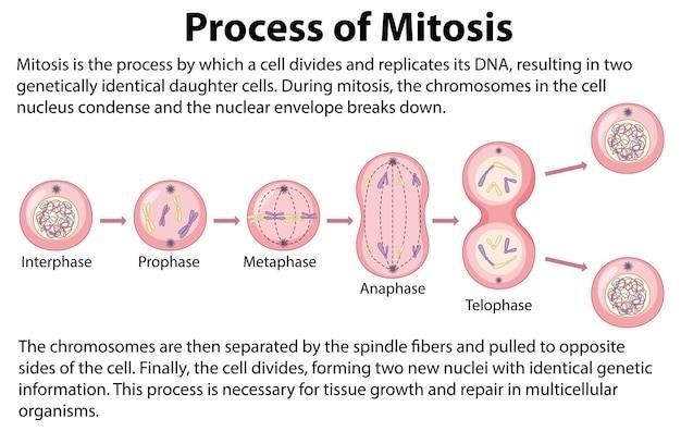
Mitosis is a fundamental process of cell division resulting in two genetically identical daughter cells from a single parent cell. This type of cell division is crucial for growth, repair, and asexual reproduction in many organisms. Mitosis ensures the precise duplication and distribution of the parent cell’s chromosomes to each daughter cell, maintaining genetic consistency. The process is characterized by a series of distinct phases⁚ prophase, metaphase, anaphase, and telophase. During prophase, chromosomes condense and become visible under a microscope; the nuclear envelope breaks down, and the mitotic spindle begins to form. In metaphase, chromosomes align at the cell’s equator, ensuring equal distribution to daughter cells. Anaphase involves the separation of sister chromatids, pulled towards opposite poles of the cell by the spindle fibers. Telophase sees the formation of two new nuclei, one at each pole, and the chromosomes begin to decondense. Cytokinesis, the division of the cytoplasm, follows telophase, resulting in two separate, genetically identical daughter cells, each with a complete set of chromosomes.
The precise regulation of mitosis is vital for maintaining genomic stability. Errors during mitosis can lead to aneuploidy (abnormal chromosome number) and contribute to genetic disorders or cancer.
Meiosis⁚ The Basics
Meiosis is a specialized type of cell division that occurs in sexually reproducing organisms, producing gametes (sperm and egg cells) with half the number of chromosomes as the parent cell. This reduction in chromosome number is essential for maintaining a constant chromosome number across generations. Unlike mitosis, meiosis involves two rounds of division⁚ Meiosis I and Meiosis II. Meiosis I is characterized by homologous chromosome pairing and recombination, leading to genetic variation in the resulting gametes. Homologous chromosomes, one inherited from each parent, pair up during prophase I, forming tetrads. Crossing over, the exchange of genetic material between homologous chromosomes, occurs during this stage, creating new combinations of alleles. In metaphase I, homologous chromosome pairs align at the metaphase plate, and in anaphase I, homologous chromosomes separate and move to opposite poles. Telophase I is followed by cytokinesis, resulting in two haploid daughter cells. Meiosis II resembles mitosis, with sister chromatids separating in anaphase II. The final result is four haploid daughter cells, each genetically unique due to crossing over and independent assortment of chromosomes.
The genetic diversity generated during meiosis is crucial for adaptation and evolution, increasing the fitness of a population in changing environments.

Comparing Mitosis and Meiosis⁚ A Chart
To effectively compare mitosis and meiosis, a chart is invaluable; This visual tool allows for a side-by-side examination of key differences and similarities. The chart should include columns for each process and rows for the following characteristics⁚ Type of cell division (somatic vs. germline), Number of daughter cells produced (two vs. four), Chromosome number of daughter cells (diploid vs. haploid), Genetic variation (none vs. significant), Purpose (growth, repair, and asexual reproduction vs. sexual reproduction), and Stages (prophase, metaphase, anaphase, telophase, and cytokinesis, noting the differences in Meiosis I and Meiosis II). A concise description of each stage in both processes should be included within the chart itself or in accompanying notes. This detailed comparison will highlight the fundamental distinctions between these two essential cellular processes. Visual aids like diagrams within the chart can greatly enhance understanding. A clear and organized chart will serve as an excellent study tool, enabling a comprehensive comparison of mitosis and meiosis.
Mitosis vs. Meiosis⁚ Key Differences
The core distinction lies in their outcomes⁚ mitosis generates two genetically identical diploid daughter cells, crucial for growth and repair within an organism. Meiosis, conversely, produces four genetically unique haploid daughter cells – gametes (sperm and egg cells) – essential for sexual reproduction. Mitosis involves a single round of cell division, while meiosis comprises two successive divisions (Meiosis I and Meiosis II). Genetic variation is absent in mitosis; daughter cells are exact replicas of the parent cell. Meiosis, however, introduces significant genetic diversity through processes like crossing over (exchange of genetic material between homologous chromosomes) and independent assortment (random distribution of chromosomes into daughter cells). This genetic shuffling is vital for evolution and adaptation within populations. Mitosis occurs in somatic cells throughout an organism’s life, whereas meiosis is confined to germ cells in reproductive organs. In summary, mitosis ensures genetic continuity within an individual, while meiosis facilitates genetic variation across generations.
Stages of Mitosis⁚ A Detailed Look
Mitosis, the process of nuclear division, unfolds in several distinct phases⁚ Prophase, characterized by chromosome condensation and the formation of the mitotic spindle; Prometaphase, where the nuclear envelope breaks down and spindle fibers attach to chromosomes; Metaphase, where chromosomes align at the cell’s equator (metaphase plate); Anaphase, marked by the separation of sister chromatids and their movement towards opposite poles; and Telophase, where chromosomes decondense, the nuclear envelope reforms, and the spindle disappears. Cytokinesis, the division of the cytoplasm, follows telophase, resulting in two genetically identical daughter cells. Each phase involves precise and tightly regulated molecular events ensuring accurate chromosome segregation. Errors during mitosis can lead to aneuploidy (abnormal chromosome number), a condition linked to various diseases, including cancer. Understanding the intricate choreography of these stages is vital for comprehending cell division and its significance in growth, development, and tissue repair.
Stages of Meiosis I⁚ A Detailed Look
Meiosis I, the first of two meiotic divisions, is a reductional division, reducing the chromosome number by half. Prophase I is unique, marked by homologous chromosome pairing (synapsis) and crossing over, a crucial process for genetic recombination. These paired chromosomes, called bivalents or tetrads, exchange genetic material, creating new combinations of alleles. Metaphase I sees bivalents aligning at the metaphase plate, with each homologous pair independently oriented. Anaphase I involves the separation of homologous chromosomes, each consisting of two sister chromatids, moving towards opposite poles. This separation is random, leading to independent assortment of chromosomes. Telophase I concludes with the formation of two haploid daughter cells, each with a reduced chromosome number but still containing duplicated chromosomes. Cytokinesis follows, completing the first meiotic division. The unique events of Prophase I—synapsis and crossing over—are responsible for generating genetic diversity in the resulting gametes, crucial for sexual reproduction and the evolution of species. These processes contribute significantly to genetic variation within populations.
Stages of Meiosis II⁚ A Detailed Look
Meiosis II closely resembles a mitotic division, but operates on haploid cells produced during Meiosis I. Prophase II begins with condensed chromosomes, each consisting of two sister chromatids, in each of the two haploid daughter cells. A new spindle apparatus forms, and the nuclear envelope breaks down. In Metaphase II, individual chromosomes align at the metaphase plate, similar to mitosis. However, unlike Meiosis I, homologous chromosomes are not paired. Anaphase II marks the separation of sister chromatids; each chromatid, now considered an individual chromosome, moves to opposite poles. This separation ensures that each daughter cell receives only one copy of each chromosome. Telophase II sees the arrival of chromosomes at the poles, followed by the reformation of nuclear envelopes and decondensation of chromosomes. Cytokinesis then divides the cytoplasm, resulting in four haploid daughter cells, each genetically unique due to the events of Meiosis I. These cells, typically gametes (sperm or egg cells), are ready for fertilization to restore the diploid chromosome number in the next generation. The process ensures genetic diversity vital for adaptation and evolution.
Genetic Variation in Meiosis
Meiosis is a cornerstone of genetic diversity, crucial for the evolution and adaptation of sexually reproducing organisms. This variation arises primarily through two mechanisms⁚ independent assortment and crossing over. Independent assortment refers to the random alignment of homologous chromosome pairs during metaphase I. The orientation of each pair is independent of others, leading to numerous possible combinations of maternal and paternal chromosomes in the resulting gametes. This randomness exponentially increases the potential genetic diversity among offspring. Crossing over, occurring during prophase I, further enhances variation. Non-sister chromatids of homologous chromosomes exchange segments of DNA at points called chiasmata. This exchange shuffles genetic material, creating recombinant chromosomes with unique combinations of alleles. The combination of independent assortment and crossing over generates a vast array of genetically distinct gametes from a single diploid cell, ensuring that offspring are not simply clones of their parents. This genetic variation is the driving force behind natural selection and the adaptation of populations to changing environments.
The Role of Mitosis in Growth and Repair
Mitosis plays a vital role in the growth and development of multicellular organisms. It’s the engine of tissue expansion, allowing organisms to increase in size from a single fertilized egg to a complex structure. Each mitotic division produces two genetically identical daughter cells, ensuring that the genetic information is faithfully replicated and passed on to every cell in the body. This precise replication is essential for maintaining the integrity and function of tissues and organs. Beyond growth, mitosis is crucial for tissue repair and regeneration. When tissues are damaged—through injury, disease, or wear and tear—mitosis provides the mechanism to replace lost or damaged cells. This process ensures the continuous renewal and maintenance of various tissues throughout an organism’s lifetime. The rate of mitosis varies significantly depending on the tissue type. Some tissues, such as skin and gut lining, undergo frequent cell division to replace rapidly worn-out cells. Others, like nerve tissue, exhibit very limited mitotic activity after development. The precise regulation of mitosis is critical for preventing uncontrolled cell growth, which can lead to cancerous tumors.
The Role of Meiosis in Sexual Reproduction
Meiosis is an essential process in sexual reproduction, responsible for generating the gametes—sperm and egg cells—that carry genetic information from parents to offspring. Unlike mitosis, which produces genetically identical daughter cells, meiosis results in four genetically unique haploid cells. This genetic variation is crucial for the survival and adaptation of species. The reduction in chromosome number from diploid (two sets of chromosomes) to haploid (one set) during meiosis ensures that when fertilization occurs, the resulting zygote will have the correct diploid number of chromosomes, maintaining genetic stability across generations. The unique genetic makeup of each gamete arises from two key events during meiosis⁚ crossing over (the exchange of genetic material between homologous chromosomes) and independent assortment (the random distribution of maternal and paternal chromosomes into daughter cells). These processes shuffle the genetic deck, creating a vast array of possible gamete combinations. This genetic diversity is the raw material upon which natural selection acts, driving evolutionary change and adaptation to environmental pressures. Without meiosis, sexual reproduction would not be possible, hindering the remarkable biodiversity seen in the natural world.
